Wednesday, October 26, 2005

Craniofacial: Medical Data Visualization

Visualizaiton tool kit (vtk4.2 and above) TCL programming with TK Widgets.

Cranio-Facial Infant Viewer- GUI

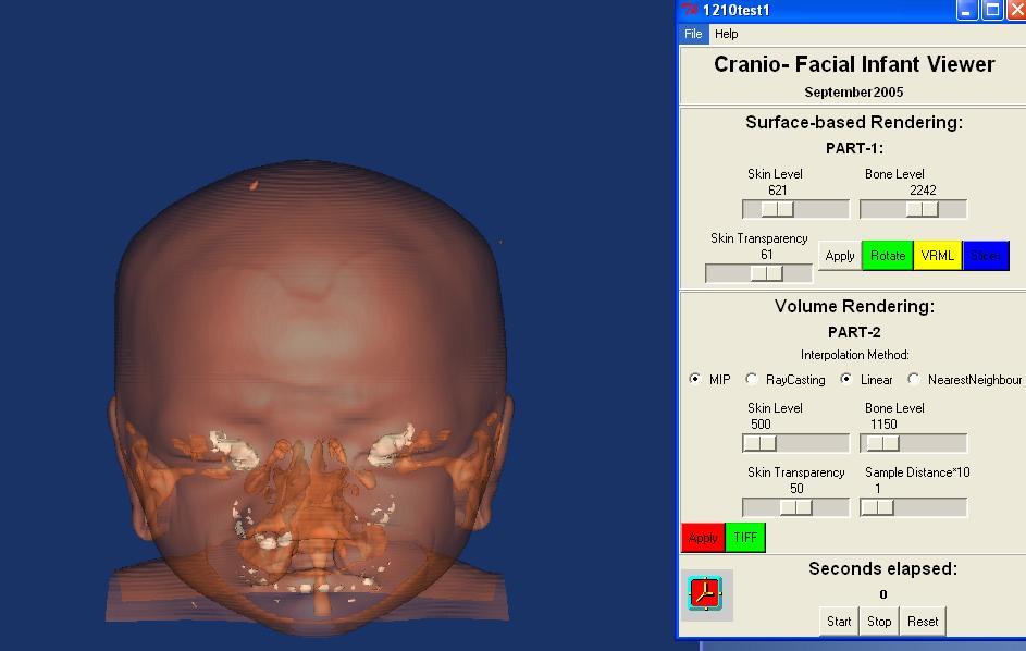
Help Files Part-1 (Step1-3) & Part-3 (Step 4-6)
1.0 User can follow the order to set isovalues:Skin Level,Bone Level,Skin Transparency then Press Apply
2.0 User can interact with ANIM button it'll create *.TIFF files for various iso values in 360 degree view takes time.
3.0 User can interact with VRML button it'll create *.wrl files using INDEXFACESET geometry.
4.0 User can follow Step1 and adjust the Sample distance and select MIP or Composite then Press Apply
5.0 User can follow Step 4 and adjust the Sample distance then select Linear or Nearest Neighbour then Press Apply
6.0 User can Press the TIFF button and can capture the current status of the image from the rendering window

7.0

2D Slice


Sagittal, Frontal, Axial buttons reset the widget to orthonormal positioning, set the horizontal slider to move the associated widget along its normal, and set the camera to face the widget
Click on Sagittal, Frontal, Axial buttons to see the different 2Ds in different Projection for X, Y and Z direction

MIP with Sample distance


MIP Raycasting for skin level 500 Bone level 1150, with linear interpolation, transparency 0 and sampling distance.

Maximum Intensity Projection (MIP)


MIP raycasting with different isovlues for skin and bone with linear interpolation.

Composite Raycasting -Linear Interpolation


Composite Raycasting for different skin and bone isovalues with linear interpolation method.

Volume Rendering

Volume Rendering:
This program will produce different volume renderings of the Infant datasets.
Volume rendering techniques have been developed to overcome problems of the accurate representation of surfaces in the isosurface techniques. Volume rendering does not use intermediate geometrical representations, in contrast to surface rendering techniques. It offers the possibility for displaying weak or fuzzy surfaces.

The term volume rendering is used to describe techniques which allow the visualization of three-dimensional data. Volume rendering is a technique for visualizing sampled functions of three spatial dimensions by computing 2-D projections of a colored semitransparent volume.
The Obvious Advantages of Volume rendering over isosurfacing.

Volume Rendering displays a dataset which convey depth information much more clearly than isosurfacing; this is not surprising since with isosurfacing we are limited to particular surface (isovalue) while with Volume Rendering we see a set of nested surfaces. But isosurfacing is much faster than volume rendering and if quick iterative results with acceptable visualization quality applied as three views (axial, frontal and sagital) on one slice, is achieving what we want then isosurfacing is good.

The major application area of volume rendering is medical imaging, where volume data is available from X-ray Computer Tomagraphy (CT) scanners. The image has given in this assignment is taken by a CT scanner. CT produce three-dimensional stacks of parallel plane images, each of which consists of an array of X-ray absorption coefficients.
The assignment dataset X-ray CT images will have a resolution of 512 * 512 * 123 bits and there will be up to 50 slides in a stack. The slides are 1-5 mm thick, and are spaced 1-5 mm apart.

Volume rendering involves the following steps: the forming of an RGBA volume from the data, reconstruction of a continuous function from this discrete data set, and projecting it onto the 2D viewing plane (the output image) from the desired point of view. An RGBA volume is a 3D four-vector data set, where the first three components are the familiar R, G, and B color components and the last component, A, represents opacity. An opacity value of 0 means totally transparent and a value of 1 means totally opaque. Behind the RGBA volume an opaque background is placed. The mapping of the data to opacity values acts as a classification of the data one is interested in. Isosurfaces can be shown by mapping the corresponding data values to almost opaque values and the rest to transparent values.

The appearance of surfaces can be improved by using shading techniques to form the RGB mapping. However, opacity can be used to see the interior of the data volume too. These interiors appear as clouds with varying density and color. A big advantage of volume rendering is that this interior information is not thrown away, so that it enables one to look at the 3D data set as a whole. Disadvantages are the difficult interpretation of the cloudy interiors and the long time, compared to surface rendering, needed to perform volume rendering.

Surface Based Rendering


Surface Based Rendering:
1- Isovalues ranges for Skin and Bone: - In order to animate the surfaces of skin and bone, we loop between different isovalues, until we determine the best isovalues range for the skin which is in our project is from 500 to1000 and from 1000 to 3000 for bone.

2- Setting up the camera object parameters: - To control the view direction, we did this by setting SetFocalPoint 0 0 0, and set position method with the parameter SetPosition 0 1 0, now also we need to control the camera movement, we set the camera parameter that determine the movement of the camera by Dolly 1.5, Dolly will move the direction of the camera along the direction of projection, moving towards the focal point.

3- Writing Multiple Tiff images: - we create image file showing the content of the render window, in order to do this we create separate button in our GUI that invoke the vtkTIFFWritter to set the input for VtkWindowToImageFilter, so finally we obtain the output Tiff image.

Data-Visualization


Introduction:
Scientific visualization utilizes computer graphics techniques to gain insight into complex and huge data sets. Medicine is one of the fields that greatly benefits from the scientific visualization techniques. Scientific visualization assists in the fields of surgical planning, beam radiation therapy, medical teaching and diagnosis.

How Visualization techniques will help the physicians in the field of Cranio-Facial Surgery?

In our project, Physicians and Doctors are concerned in the medical studies of Cranio-Facial deformities, for a sample of Malaysians infants, they are trying finally to do corrective surgery (more like cosmetic surgery on certain part of cranio facial), so they need assistance from some visualization techniques to help them for better studying and analysis for the Cranio-Facial deformities before they do the surgery for the patient infants.

We obtained our dataset for this project as medical images scanned with modern CT and MRI scanner, from the Medical Campus in Kelantan at the University Sains Malaysia.

We are trying to visualize this dataset with some appropriate volumetric visualization techniques, these techniques varying from 1D scalar techniques as snapshot in Tiff image format or VRML format, to 2D Scalar techniques as 2Ds slices (axial, frontal and sagittal) this techniques know as SLICING through the 3D volume by reducing it to 2D problem, finally we apply different 3D visualization techniques like surface extraction since we extracted different surface for each skin and bone according to constant values for each of them, also ray casting with composite and MIP techniques is used in our project as volume visualization techniques.

We have chosen the VTK 4.2 with TCL programming as the visualization language in our project, because the VTK utilizes the pipeline model to describe the flow of data, and how the core of the classes interfaces with each other. Sources start the visualization pipeline, filter operates on the data as enrichment process that passes through it, a Mapper objects terminates the data flow and contains the final representation of the data that will be passes to a render. If a Mapper writes the data to a file it is referred as a writer object. This makes visualization with VTK straightforward and simple.

Thursday, October 20, 2005

Create an MPEG Movie

Animating a series of Tiff flies allowed to create a GIF file. This File tells us about various isovalues are being changes to visualize the various features in the volume data . To make this GIF there are 1500 TIFF files are beign used.

Wednesday, October 12, 2005

Cranio-Facial -Final Stages 95%

Tuesday, September 27, 2005

Cranio-Facial Visualization with GUI


Working with DICOM data, mapping the image files into Surface-based Rendering and Volume-Rendering. The main usage of the GUI it to help the medical doctor's to change the parameters for skin and bone.

Visualizing the volume data with the Interpolation methods such as MIP and Composite. By setting up our own lookuptable to show the distinctively the various features in the 2D slice (soft tissue, skin, bone, arteries etc.,)


Hey, this is just 70% of the work. I have to finish an another 30%.

Any comments?

Monday, September 19, 2005

Comment from Rough Jade

Hey Guys,
This is just only 50% of the work. I hope i can finish it soon.
Note:
Software platform : vtk4.2 with Tcl/Tk (Tickle)
Operating System : Windows Xp SpII

Volume Rendering, Surface Rendering; Transparency


The Focus is on employing surface-based rendering techniques using vtkWindowToImageFiler, vtkVolume16Reader, vtkTiffWriter, vtkCamera, vtkRayCastMapper etc., This is to help the physician to iterate through the slices interactively.

Wednesday, September 14, 2005

Isosurface Visualization

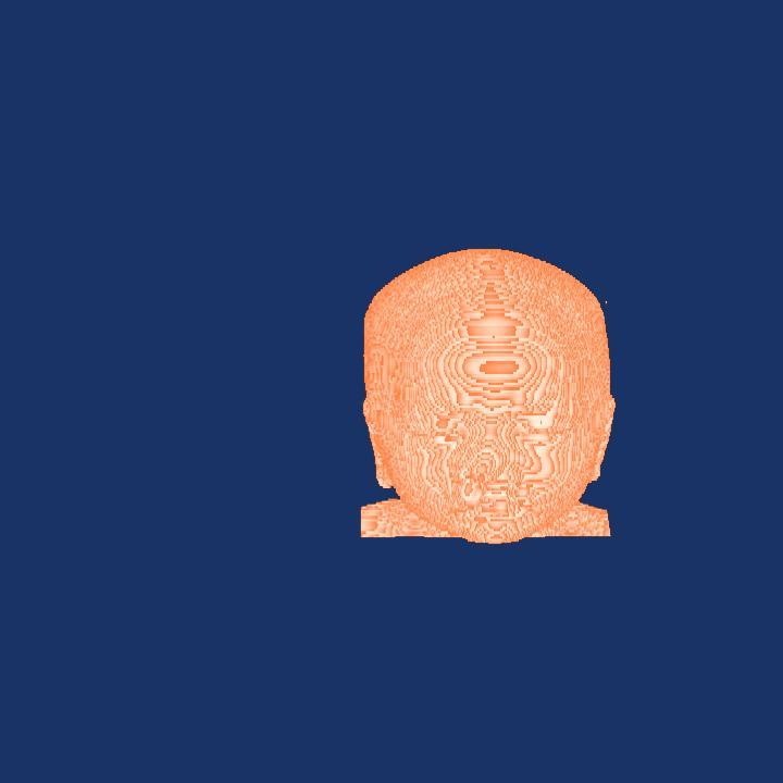
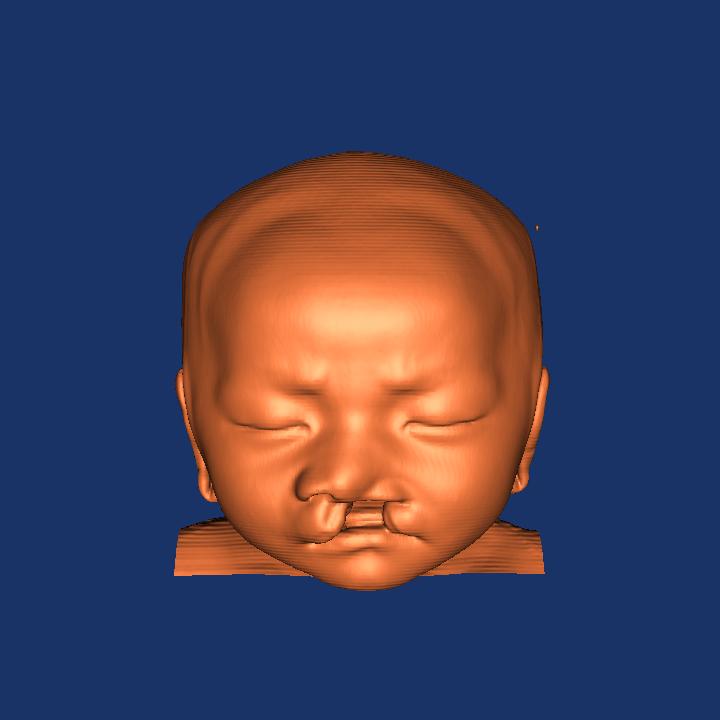
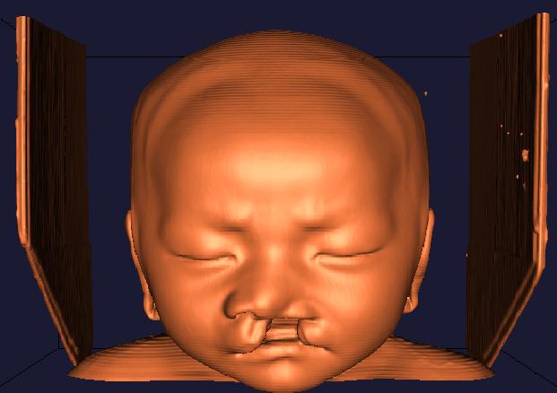
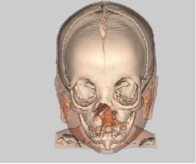
Here is the isosurface visualization and are fully implemented and working properly. Two isosurfaces are provided - One for skin and one for bone. The transparency of the skin is adjustable from 0 to 100%, and the levels which "define" skin and bone are also adjustable within a reasonable range. Resolution is selectable from 32 to 256 in powers of 2.

Sunday, September 04, 2005

Cranio-Facial


Cronio-facial is a medical term refering to the head(skull) and face of the human body. Medical study on cranio-facial are abound, rancing from grey matter investigations, nerves intricacies, speech and hearing organs complexities, and finally leading toward anatomical and skeletal deformities such as cleft and various protrusions.